| | | | 實用人體解剖彩色圖譜(第3版)(精) | | 該商品所屬分類:醫學 -> 醫技學 | | 【市場價】 | 768-1113元 | | 【優惠價】 | 480-696元 | | 【介質】 | book | | 【ISBN】 | 9787117258111 | | 【折扣說明】 | 一次購物滿999元台幣免運費+贈品
一次購物滿2000元台幣95折+免運費+贈品
一次購物滿3000元台幣92折+免運費+贈品
一次購物滿4000元台幣88折+免運費+贈品
| | 【本期贈品】 | ①優質無紡布環保袋,做工棒!②品牌簽字筆 ③品牌手帕紙巾
|
|
| 版本 | 正版全新電子版PDF檔 | | 您已选择: | 正版全新 | 溫馨提示:如果有多種選項,請先選擇再點擊加入購物車。*. 電子圖書價格是0.69折,例如了得網價格是100元,電子書pdf的價格則是69元。
*. 購買電子書不支持貨到付款,購買時選擇atm或者超商、PayPal付款。付款後1-24小時內通過郵件傳輸給您。
*. 如果收到的電子書不滿意,可以聯絡我們退款。謝謝。 | | | |
| | 內容介紹 | |

-
出版社:人民衛生
-
ISBN:9787117258111
-
作者:編者:羊惠君
-
頁數:278
-
出版日期:2018-03-01
-
印刷日期:2018-03-01
-
包裝:精裝
-
開本:16開
-
版次:3
-
印次:16
-
字數:462千字
-
-
★實物圖譜的**者,已是10萬+醫學生的選擇!
★書中通過實物展現人體結構,局解和繫解兼顧。
★本圖譜按照醫學院校人體解剖學課程進度安排章節內容,方便醫學生學習使用。
-
本圖譜按照醫學院校人體解剖學課程進度安排章節內容,方便醫學生學習使用。第1章為緒論,簡要介紹人體各繫統的組成。第2章至第8章依次為下肢、上肢、胸、腹、盆與會陰、背、頭與頸。第9章為中樞神經繫統,集中介紹腦、脊髓的結構和功能聯繫。
◎本圖譜為充分顯示結構復雜的局部器官,使用了381件精心制作的實物標本、22幅手繪示意圖、4張活體彩色照片和1張牙齒的X線片。標本制備細致,照片質量高,結構標注詳盡。學生在解剖學實驗室用本圖譜對照觀察解剖出來的結構器官,可很快準確理解他們所應掌握的解剖學理論和知識。本圖譜按照醫學院校人體解剖學課程進度安排章節內容,方便醫學生學習使用。第1章為緒論,簡要介紹人體各繫統的組成。第2章至第8章依次為下肢、上肢、胸、腹、盆與會陰、背、頭與頸。第9章為中樞神經繫統,集中介紹腦、脊髓的結構和功能聯繫。
◎本圖譜為充分顯示結構復雜的局部器官,使用了381件精心制作的實物標本、22幅手繪示意圖、4張活體彩色照片和1張牙齒的X線片。標本制備細致,照片質量高,結構標注詳盡。學生在解剖學實驗室用本圖譜對照觀察解剖出來的結構器官,可很快準確理解他們所應掌握的解剖學理論和知識。
-
羊惠君,人體解剖與組織胚胎學教授,博士生導師,獲國務院政府特殊津貼,四川省學術和技術帶頭人,四川省教學名師, 中國解剖學會理事,《解剖學報》編委,《解剖學雜志》編委,四川省解剖學會理事長,《四川解剖學雜志》主編,四川省精品課程《人體解剖學》課程負責人,四川省重點教育廳實驗室\"\"干細胞應用研究中心\"\"主任委員,四川省重點學科《人體解剖學與組織胚胎學》學術帶頭人。
-
目錄CONTENTS
第1章 緒論 Chapter 1 Introduction1(—10)
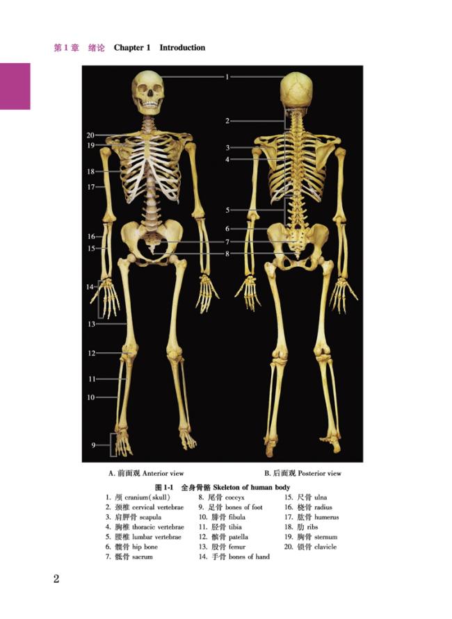
圖1-1 全身骨骼 Skeleton of human body2
圖1-2 骨的形態 Shape of bones3
圖1-3 骨的構造(股骨上端剖面) Structure of bone(section of upper part of femur)3
圖1-4 骨的類型 Type of bones3
圖1-5 骨骼肌形態 Shape of skeletal muscles4
圖1-6 呼吸繫統(前面觀) Respiratory system(anterior view)5
圖1-7 消化繫統(前面觀) Digestive system(anterior view)6
圖1-8 腸壁的層次 Layers of intestinal wall6
圖1-9 男性膀胱和生殖繫統(前面觀) Male urinary bladder and reproductive system(anterior view)7
圖1-10 女性生殖繫統(前面觀) Female reproductive system(anterior view)8
圖1-11 男性內分泌器官(前面觀) Endocrine organs of male(anterior view)9
圖1-12 神經繫統(前面觀) Nervous system(anterior view)10
第2章 下肢 Chapter 2 Lower Limb11(—43)
圖2-1 右側髖骨 Right hip bone12
圖2-2 右側嬰兒髖骨(外面觀) Right infant hip bone(external view)12
圖2-3 右側股骨 Right femur13
圖2-4 右側髕骨 Right patella14
圖2-5 右側脛骨與腓骨 Right tibia and fibula14
圖2-6 右側足骨 Bones of right foot15
圖2-7 右側足縱弓 Longitudinal arches of right foot16
圖2-8 骨盆的連接 Joints of pelvis17
圖2-9 右髖關節 Right hip joint18
圖2-10 右膝關節 Right knee joint19
圖2-11 剖開的右膝關節 Dissected right knee joint20
圖2-12 右側膝關節半月板(上面觀) Meniscuses of right knee joint(superior view)21
圖2-13 右側膝關節矢狀切面 Sagittal section through right knee joint21
圖2-14 右側小腿骨連接(前面觀) Joints of right leg (anterior view)22
圖2-15 右側踝關節周圍的韌帶 Ligaments around right a目錄CONTENTS
第1章 緒論 Chapter 1 Introduction1(—10)
圖1-1 全身骨骼 Skeleton of human body2
圖1-2 骨的形態 Shape of bones3
圖1-3 骨的構造(股骨上端剖面) Structure of bone(section of upper part of femur)3
圖1-4 骨的類型 Type of bones3
圖1-5 骨骼肌形態 Shape of skeletal muscles4
圖1-6 呼吸繫統(前面觀) Respiratory system(anterior view)5
圖1-7 消化繫統(前面觀) Digestive system(anterior view)6
圖1-8 腸壁的層次 Layers of intestinal wall6
圖1-9 男性膀胱和生殖繫統(前面觀) Male urinary bladder and reproductive system(anterior view)7
圖1-10 女性生殖繫統(前面觀) Female reproductive system(anterior view)8
圖1-11 男性內分泌器官(前面觀) Endocrine organs of male(anterior view)9
圖1-12 神經繫統(前面觀) Nervous system(anterior view)10
第2章 下肢 Chapter 2 Lower Limb11(—43)
圖2-1 右側髖骨 Right hip bone12
圖2-2 右側嬰兒髖骨(外面觀) Right infant hip bone(external view)12
圖2-3 右側股骨 Right femur13
圖2-4 右側髕骨 Right patella14
圖2-5 右側脛骨與腓骨 Right tibia and fibula14
圖2-6 右側足骨 Bones of right foot15
圖2-7 右側足縱弓 Longitudinal arches of right foot16
圖2-8 骨盆的連接 Joints of pelvis17
圖2-9 右髖關節 Right hip joint18
圖2-10 右膝關節 Right knee joint19
圖2-11 剖開的右膝關節 Dissected right knee joint20
圖2-12 右側膝關節半月板(上面觀) Meniscuses of right knee joint(superior view)21
圖2-13 右側膝關節矢狀切面 Sagittal section through right knee joint21
圖2-14 右側小腿骨連接(前面觀) Joints of right leg (anterior view)22
圖2-15 右側踝關節周圍的韌帶 Ligaments around right a章 盆與會陰 Chapter 6 Pelvis and Perineum129(—150)
圖6-1 男性骨盆上口(上面觀) Superior pelvic aperture of male(superior view)130
圖6-2 男性骨盆下口(下面觀) Inferior pelvic aperture of male(inferior view)130
圖6-3 女性骨盆上口(上面觀) Superior pelvic aperture of female(superior view)131
圖6-4 女性骨盆下口(下面觀) Inferior pelvic aperture of female(inferior view)131
圖6-5 男性肛提肌(上面觀) Levator ani of male(superior view)132
圖6-6 男性盆部(上面觀) Pelvis of male(superior view)133
圖6-7 男性盆部正中矢狀面(右側面觀) Median sagittal section of male pelvis(right lateral view)134
圖6-8 男性盆腔(右側面觀) Pelvic cavity of male(right lateral view)134
圖6-9 男性盆部動脈(上面觀) Arteries of male pelvis(superior view)135
圖6-10 男性盆部動脈(右側面觀) Arteries of male pelvis(right lateral view)135
圖6-11 男性盆部神經(上面觀) Nerves of male pelvis(superior view)136
圖6-12 男性膀胱(內面觀) Male urinary bladder(internal view)137
圖6-13 前列腺和精囊(後面觀) Prostate and seminal vesicle(posterior view)137
圖6-14 男性膀胱底(後面觀) Fundus of male urinary bladder(posterior view)138
圖6-15 直腸(內面觀) Rectum(internal view)139
圖6-16 膀胱、前列腺和精囊腺(右側面觀) Urinary bladder, prostate and seminal vesicle(right lateral view)139
圖6-17 前列腺橫斷面 Transverse section of prostate139
圖6-18 女性盆部正中矢狀剖面(右側面觀) Median sagittal section of female pelvis(right lateral view)140
圖6-19 卵巢和子宮(前面觀,膀胱和左側半盆腔腹膜已移除) Ovary and uterus(anterior view, urinary bl章 盆與會陰 Chapter 6 Pelvis and Perineum129(—150)
圖6-1 男性骨盆上口(上面觀) Superior pelvic aperture of male(superior view)130
圖6-2 男性骨盆下口(下面觀) Inferior pelvic aperture of male(inferior view)130
圖6-3 女性骨盆上口(上面觀) Superior pelvic aperture of female(superior view)131
圖6-4 女性骨盆下口(下面觀) Inferior pelvic aperture of female(inferior view)131
圖6-5 男性肛提肌(上面觀) Levator ani of male(superior view)132
圖6-6 男性盆部(上面觀) Pelvis of male(superior view)133
圖6-7 男性盆部正中矢狀面(右側面觀) Median sagittal section of male pelvis(right lateral view)134
圖6-8 男性盆腔(右側面觀) Pelvic cavity of male(right lateral view)134
圖6-9 男性盆部動脈(上面觀) Arteries of male pelvis(superior view)135
圖6-10 男性盆部動脈(右側面觀) Arteries of male pelvis(right lateral view)135
圖6-11 男性盆部神經(上面觀) Nerves of male pelvis(superior view)136
圖6-12 男性膀胱(內面觀) Male urinary bladder(internal view)137
圖6-13 前列腺和精囊(後面觀) Prostate and seminal vesicle(posterior view)137
圖6-14 男性膀胱底(後面觀) Fundus of male urinary bladder(posterior view)138
圖6-15 直腸(內面觀) Rectum(internal view)139
圖6-16 膀胱、前列腺和精囊腺(右側面觀) Urinary bladder, prostate and seminal vesicle(right lateral view)139
圖6-17 前列腺橫斷面 Transverse section of prostate139
圖6-18 女性盆部正中矢狀剖面(右側面觀) Median sagittal section of female pelvis(right lateral view)140
圖6-19 卵巢和子宮(前面觀,膀胱和左側半盆腔腹膜已移除) Ovary and uterus(anterior view, urinary bladder and left half of peritoneum has been removed)141
圖6-20 女性盆部動脈(上面觀) Arteries of female pelvis(superior view)142
圖6-21 男性會陰淺隙和坐骨肛門窩(Ⅰ)(下面觀) Superficial perineal space and ischioanal fossa of male(Ⅰ)(inferior view)143
圖6-22 男性會陰淺隙和坐骨肛門窩(Ⅱ)(下面觀) Superficial perineal space and ischioanal fossa of male(Ⅱ)(inferior view)144
圖6-23 男性會陰深隙和坐骨肛門窩(下面觀) Deep perineal space and ischioanal fossa of male(inferior view)145
圖6-24 睪丸和精索被膜(前面觀) Testis and tunicae of spermatic cord(anterior view)146
圖6-25 陰囊層次(前面觀) Layers of scrotum(anterior view)146
圖6-26 陰莖層次(背側觀) Layers of penis(dorsal view)147
圖6-27 陰莖勃起組織(腹側觀) Erectile tissues of penis(ventral view)147
圖6-28 女性外生殖器 External genital organs of female148
圖6-29 女性會陰淺隙和坐骨肛門窩(Ⅰ)(下面觀) Superficial perineal space and ischioanal fossa of female(Ⅰ)(inferior view)149
圖6-30 女性會陰淺隙和坐骨肛門窩(Ⅱ)(下面觀) Superficial perineal space and ischioanal fossa of female(Ⅱ)(inferior view)150
第7章 背 Chapter 7 Back151(—161)
圖7-1 脊柱 Vertebral column152
圖7-2 寰椎 Atlas153
圖7-3 樞椎(上面觀) Axis(superior view)153
圖7-4 頸椎(上面觀) A cervical vertebra(superior view)153
圖7-5 胸椎 A thoracic vertebra154
圖7-6 腰椎 A lumbar vertebra154
圖7-7 骶骨和尾骨(前面觀) Sacrum and coccyx(anterior view)155
圖7-8 骶骨和尾骨(後面觀) Sacrum and coccyx(posterior view)155
圖7-9 椎骨間連接Intervertebral jointsadder and left half of peritoneum has been removed)141
圖6-20 女性盆部動脈(上面觀) Arteries of female pelvis(superior view)142
圖6-21 男性會陰淺隙和坐骨肛門窩(Ⅰ)(下面觀) Superficial perineal space and ischioanal fossa of male(Ⅰ)(inferior view)143
圖6-22 男性會陰淺隙和坐骨肛門窩(Ⅱ)(下面觀) Superficial perineal space and ischioanal fossa of male(Ⅱ)(inferior view)144
圖6-23 男性會陰深隙和坐骨肛門窩(下面觀) Deep perineal space and ischioanal fossa of male(inferior view)145
圖6-24 睪丸和精索被膜(前面觀) Testis and tunicae of spermatic cord(anterior view)146
圖6-25 陰囊層次(前面觀) Layers of scrotum(anterior view)146
圖6-26 陰莖層次(背側觀) Layers of penis(dorsal view)147
圖6-27 陰莖勃起組織(腹側觀) Erectile tissues of penis(ventral view)147
圖6-28 女性外生殖器 External genital organs of female148
圖6-29 女性會陰淺隙和坐骨肛門窩(Ⅰ)(下面觀) Superficial perineal space and ischioanal fossa of female(Ⅰ)(inferior view)149
圖6-30 女性會陰淺隙和坐骨肛門窩(Ⅱ)(下面觀) Superficial perineal space and ischioanal fossa of female(Ⅱ)(inferior view)150
第7章 背 Chapter 7 Back151(—161)
圖7-1 脊柱 Vertebral column152
圖7-2 寰椎 Atlas153
圖7-3 樞椎(上面觀) Axis(superior view)153
圖7-4 頸椎(上面觀) A cervical vertebra(superior view)153
圖7-5 胸椎 A thoracic vertebra154
圖7-6 腰椎 A lumbar vertebra154
圖7-7 骶骨和尾骨(前面觀) Sacrum and coccyx(anterior view)155
圖7-8 骶骨和尾骨(後面觀) Sacrum and coccyx(posterior view)155
圖7-9 椎骨間連接Intervertebral joints156
圖7-10 顱骨與椎骨連接 Joints of the skull and vertebra157
圖7-11 椎管(胸段,上面觀) Vertebral canal(in thoracic region,superior view)158
圖7-12 椎間孔(右側面觀) Intervertebral foramen(right lateral view)158
圖7-13 背部淺層肌(後面觀) Superficial muscles of back(posterior view)159
圖7-14 背部深層肌(後面觀) Deep muscles of back(posterior view)160
圖7-15 枕下三角(後面觀) Suboccipital triangle(posterior view)161
第8章 頭與頸 Chapter 8 Head and Neck162(—218)
圖8-1 顱(前面觀) Skull(anterior view)163
圖8-2 顱(右側面觀) Skull(right lateral view)164
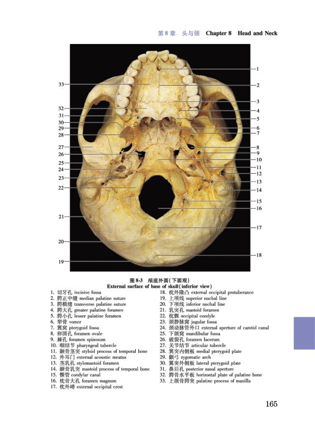
圖8-3 顱底外面(下面觀) External surface of base of skull(inferior view)165
圖8-4 顱底內面(上面觀) Internal surface of base of skull(superior view)166
圖8-5 顱頂外面(上面觀) External surface of cranial vault(superior view)167
圖8-6 新生兒顱 Skull of neonate167
圖8-7 右眶(前面觀) Right orbit(anterior view)168
圖8-8 舌骨(上面觀) Hyoid bone(superior view)168
圖8-9 下頜骨 Mandible169
圖8-10 蝶骨 Sphenoid bone170
圖8-11 右顳骨 Right temporal bone171
圖8-12 額骨 Frontal bone172
圖8-13 篩骨 Ethmoid172
圖8-14 枕骨 Occipital bone173
圖8-15 右上頜骨 Right maxilla174
圖8-16 右耳廓 Right auricle175
圖8-17 聽小骨 Auditory ossicles175
圖8-18 骨迷路 Bony labyrinth175
圖8-19 左顳下頜關節 Left temporomandibular joint176
圖8-20 頭皮 Scalp176
圖8-21 頭皮的血管(上面觀) Blood vessels of scalp(superior view)177
圖8-22 表情肌(右前外側面觀) Muscles of facial expresion(right anterolateral view)178
圖8-23 頭156
圖7-10 顱骨與椎骨連接 Joints of the skull and vertebra157
圖7-11 椎管(胸段,上面觀) Vertebral canal(in thoracic region,superior view)158
圖7-12 椎間孔(右側面觀) Intervertebral foramen(right lateral view)158
圖7-13 背部淺層肌(後面觀) Superficial muscles of back(posterior view)159
圖7-14 背部深層肌(後面觀) Deep muscles of back(posterior view)160
圖7-15 枕下三角(後面觀) Suboccipital triangle(posterior view)161
第8章 頭與頸 Chapter 8 Head and Neck162(—218)
圖8-1 顱(前面觀) Skull(anterior view)163
圖8-2 顱(右側面觀) Skull(right lateral view)164
圖8-3 顱底外面(下面觀) External surface of base of skull(inferior view)165
圖8-4 顱底內面(上面觀) Internal surface of base of skull(superior view)166
圖8-5 顱頂外面(上面觀) External surface of cranial vault(superior view)167
圖8-6 新生兒顱 Skull of neonate167
圖8-7 右眶(前面觀) Right orbit(anterior view)168
圖8-8 舌骨(上面觀) Hyoid bone(superior view)168
圖8-9 下頜骨 Mandible169
圖8-10 蝶骨 Sphenoid bone170
圖8-11 右顳骨 Right temporal bone171
圖8-12 額骨 Frontal bone172
圖8-13 篩骨 Ethmoid172
圖8-14 枕骨 Occipital bone173
圖8-15 右上頜骨 Right maxilla174
圖8-16 右耳廓 Right auricle175
圖8-17 聽小骨 Auditory ossicles175
圖8-18 骨迷路 Bony labyrinth175
圖8-19 左顳下頜關節 Left temporomandibular joint176
圖8-20 頭皮 Scalp176
圖8-21 頭皮的血管(上面觀) Blood vessels of scalp(superior view)177
圖8-22 表情肌(右前外側面觀) Muscles of facial expresion(right anterolateral view)178
圖8-23 頭頸淺結構(右外側面觀) Superficial structures of head and neck(right lateral view)178
圖8-24 面神經(Ⅶ)面部分支(右外側面觀) Branches of facial nerve(Ⅶ)on face(right lateral view)179
圖8-25 頸外動脈及其分支(右外側面觀) External carotid artery and its branches(right lateral view)180
圖8-26 頭頸靜脈(右外側面觀) Veins of head and neck(right lateral view)181
圖8-27 頭頸深結構(右外側面觀) Deep structures of head and neck(right lateral view)182
圖8-28 咀嚼肌(右外側面觀) Muscles of mastication(right lateral view)183
圖8-29 三叉神經(Ⅴ) Trigeminal nerve(Ⅴ)184
圖8-30 三叉神經(Ⅴ)(右外側面觀) Trigeminal nerve(Ⅴ)(right lateral view)185
圖8-31 面神經(Ⅶ)和舌咽神經(Ⅸ)的副交感纖維分布模式圖 Diagram for distribution of parasympathetic fibers in facial and glossharyngeal nerve(Ⅶ and Ⅸ)185
圖8-32 大唾液腺(右外側面觀) Large salivary glands(right lateral view)186
圖8-33 舌神經和舌下神經(Ⅻ)(右外側面觀) Lingual nerve and hypoglossal nerve(Ⅻ)(right lateral view)186
圖8-34 舌 Tongue187
圖8-35 口腔底肌 Muscular floor of oral cavity188
圖8-36 上頜恆、乳牙(下面觀) Upper permanent and deciduous teeth(inferior view)188
圖8-37 下頜恆、乳牙(上面觀) Lower permanent and deciduous teeth(superior view)188
圖8-38 恆牙 Permanent teeth189
圖8-39 全頜曲面斷層攝影 Orthopantomography of permanent teeth189
圖8-40 牙頰舌縱切面模式圖 Diagram of a tooth sectioned in buccolingual longitudinal plane189
圖8-41 咽峽(前面觀) Pharyngeal isthmus(anterior view)190
圖8-42 咽腔(後面觀,咽後壁已被切開並頸淺結構(右外側面觀) Superficial structures of head and neck(right lateral view)178
圖8-24 面神經(Ⅶ)面部分支(右外側面觀) Branches of facial nerve(Ⅶ)on face(right lateral view)179
圖8-25 頸外動脈及其分支(右外側面觀) External carotid artery and its branches(right lateral view)180
圖8-26 頭頸靜脈(右外側面觀) Veins of head and neck(right lateral view)181
圖8-27 頭頸深結構(右外側面觀) Deep structures of head and neck(right lateral view)182
圖8-28 咀嚼肌(右外側面觀) Muscles of mastication(right lateral view)183
圖8-29 三叉神經(Ⅴ) Trigeminal nerve(Ⅴ)184
圖8-30 三叉神經(Ⅴ)(右外側面觀) Trigeminal nerve(Ⅴ)(right lateral view)185
圖8-31 面神經(Ⅶ)和舌咽神經(Ⅸ)的副交感纖維分布模式圖 Diagram for distribution of parasympathetic fibers in facial and glossharyngeal nerve(Ⅶ and Ⅸ)185
圖8-32 大唾液腺(右外側面觀) Large salivary glands(right lateral view)186
圖8-33 舌神經和舌下神經(Ⅻ)(右外側面觀) Lingual nerve and hypoglossal nerve(Ⅻ)(right lateral view)186
圖8-34 舌 Tongue187
圖8-35 口腔底肌 Muscular floor of oral cavity188
圖8-36 上頜恆、乳牙(下面觀) Upper permanent and deciduous teeth(inferior view)188
圖8-37 下頜恆、乳牙(上面觀) Lower permanent and deciduous teeth(superior view)188
圖8-38 恆牙 Permanent teeth189
圖8-39 全頜曲面斷層攝影 Orthopantomography of permanent teeth189
圖8-40 牙頰舌縱切面模式圖 Diagram of a tooth sectioned in buccolingual longitudinal plane189
圖8-41 咽峽(前面觀) Pharyngeal isthmus(anterior view)190
圖8-42 咽腔(後面觀,咽後壁已被切開並推向兩側) Pharyngeal cavity(posterior view, posterior wall of pharynx has been cut and reflected laterally on either side)190
圖8-43 鼻的骨骼 Bony and cartilaginous skeleton of nose191
圖8-44 左側鼻旁竇的開口(內側面觀) Orifices of left paranasal sinuses(medial view)191
圖8-45 鼻腔左外側壁 Left lateral wall of nasal cavity192
圖8-46 右淚器(前面觀) Right lacrimal apparatus(anterior view)193
圖8-47 右眶隔(前面觀) Right orbital septum(anterior view)193
圖8-48 右眼球外肌(外側面觀) Right extraocular muscles(lateral view)194
圖8-49 右眼球外肌(上面觀) Right extraocular muscles(superior view)194
圖8-50 右眶內結構(右外側面觀) Contents in right orbit(right lateral view)195
圖8-51 右眼眶內容(上面觀) Contents in right orbital(superior view)195
圖8-52 右眼球水平切面模式圖(下面觀) Diagram for horizontal section of left eyeball(inferior view)196
圖8-53 眼球前半模式圖(後面觀) Diagram for anterior half of eyeball(posterior view)196
圖8-54 右眼眶水平斷面(上面觀) Horizontal section through right orbit(superior view)197
圖8-55 右眼球及眶矢狀切面(外側面觀) Sagittal section through right eyeball and orbital cavity(lateral view)197
圖8-56 右眼球前半(後面觀) Anterior half of right eyeball(posterior view)198
圖8-57 眼底血管(活體眼底鏡照片) Fundus photograph of right eye198
圖8-58 上眼瞼切片 Section of upper eyelid198
圖8-59 右前庭蝸器(上面觀) Right vestibulocochlear organ(superior view)199
圖8-60 右前庭蝸器(前面觀) Right vestibulocochlear organ(anterior 推向兩側) Pharyngeal cavity(posterior view, posterior wall of pharynx has been cut and reflected laterally on either side)190
圖8-43 鼻的骨骼 Bony and cartilaginous skeleton of nose191
圖8-44 左側鼻旁竇的開口(內側面觀) Orifices of left paranasal sinuses(medial view)191
圖8-45 鼻腔左外側壁 Left lateral wall of nasal cavity192
圖8-46 右淚器(前面觀) Right lacrimal apparatus(anterior view)193
圖8-47 右眶隔(前面觀) Right orbital septum(anterior view)193
圖8-48 右眼球外肌(外側面觀) Right extraocular muscles(lateral view)194
圖8-49 右眼球外肌(上面觀) Right extraocular muscles(superior view)194
圖8-50 右眶內結構(右外側面觀) Contents in right orbit(right lateral view)195
圖8-51 右眼眶內容(上面觀) Contents in right orbital(superior view)195
圖8-52 右眼球水平切面模式圖(下面觀) Diagram for horizontal section of left eyeball(inferior view)196
圖8-53 眼球前半模式圖(後面觀) Diagram for anterior half of eyeball(posterior view)196
圖8-54 右眼眶水平斷面(上面觀) Horizontal section through right orbit(superior view)197
圖8-55 右眼球及眶矢狀切面(外側面觀) Sagittal section through right eyeball and orbital cavity(lateral view)197
圖8-56 右眼球前半(後面觀) Anterior half of right eyeball(posterior view)198
圖8-57 眼底血管(活體眼底鏡照片) Fundus photograph of right eye198
圖8-58 上眼瞼切片 Section of upper eyelid198
圖8-59 右前庭蝸器(上面觀) Right vestibulocochlear organ(superior view)199
圖8-60 右前庭蝸器(前面觀) Right vestibulocochlear organ(anterior view)199
圖8-61 右前庭蝸器模式圖(前面觀) Diagram of right vestibulocochlear organ(anterior view)200
圖8-62 右鼓室外側壁模式圖(內側面觀) Diagram for lateral wall of right tympanic cavity(medial view)200
圖8-63 右內耳道底模式圖(內側面觀) Diagram for bottom of right internal acoustic meatus(medial view)201
圖8-64 右鼓膜模式圖(外側觀) Diagram for right tympanic membrane(lateral view)201
圖8-65 耳蝸斷面模式圖 Diagram for section through cochlea201
圖8-66 右骨迷路內面模式圖 Diagram for interior of right bony labyrinth202
圖8-67 右膜迷路模式圖 Diagram for right membranous labyrinty202
圖8-68 頭部橫斷面(經樞椎齒突) Transverse section through head at level of dens of axis203
圖8-69 頸闊肌(前面觀) Platysma(anterior view)204
圖8-70 頸闊肌(右前外側面觀) Platysma(right anterolateral view)204
圖8-71 頸淺靜脈(Ⅰ)(前面觀) Superficial veins of neck(Ⅰ)(anterior view)205
圖8-72 頸淺靜脈(Ⅱ)(前面觀) Superficial veins of neck(Ⅱ)(anterior view)205
圖8-73 頸叢和頸淺靜脈(右外側面觀) Cervical plexus and superficial jugular veins(right lateral view)206
圖8-74 頸深筋膜淺層(右外側面觀) Superficial layer of cervical deep fascia(right lateral view)206
圖8-75 頸部三角(右前外側面觀) Triangles of neck(right anterolateral view)207
圖8-76 頸前區肌(前面觀) Muscles of anterior cervical region(anterior view)207
圖8-77 頸叢分支(右外側面觀) Branches of cervical plexus(right lateral view)208
圖8-78 甲狀腺區(前面觀) Region of thyroid gland(anterior view)208
圖8-79 頸部淺層結構(右前外側view)199
圖8-61 右前庭蝸器模式圖(前面觀) Diagram of right vestibulocochlear organ(anterior view)200
圖8-62 右鼓室外側壁模式圖(內側面觀) Diagram for lateral wall of right tympanic cavity(medial view)200
圖8-63 右內耳道底模式圖(內側面觀) Diagram for bottom of right internal acoustic meatus(medial view)201
圖8-64 右鼓膜模式圖(外側觀) Diagram for right tympanic membrane(lateral view)201
圖8-65 耳蝸斷面模式圖 Diagram for section through cochlea201
圖8-66 右骨迷路內面模式圖 Diagram for interior of right bony labyrinth202
圖8-67 右膜迷路模式圖 Diagram for right membranous labyrinty202
圖8-68 頭部橫斷面(經樞椎齒突) Transverse section through head at level of dens of axis203
圖8-69 頸闊肌(前面觀) Platysma(anterior view)204
圖8-70 頸闊肌(右前外側面觀) Platysma(right anterolateral view)204
圖8-71 頸淺靜脈(Ⅰ)(前面觀) Superficial veins of neck(Ⅰ)(anterior view)205
圖8-72 頸淺靜脈(Ⅱ)(前面觀) Superficial veins of neck(Ⅱ)(anterior view)205
圖8-73 頸叢和頸淺靜脈(右外側面觀) Cervical plexus and superficial jugular veins(right lateral view)206
圖8-74 頸深筋膜淺層(右外側面觀) Superficial layer of cervical deep fascia(right lateral view)206
圖8-75 頸部三角(右前外側面觀) Triangles of neck(right anterolateral view)207
圖8-76 頸前區肌(前面觀) Muscles of anterior cervical region(anterior view)207
圖8-77 頸叢分支(右外側面觀) Branches of cervical plexus(right lateral view)208
圖8-78 甲狀腺區(前面觀) Region of thyroid gland(anterior view)208
圖8-79 頸部淺層結構(右前外側觀) Superficial layer of structures of neck(right anterolateral view)209
圖8-80 頸部中層結構(右前外側面觀) Middle layer of structures of neck(right anterolateral view)210
圖8-81 頸部深層結構(右前外側面觀) Deep layer of structures of neck(right anterolateral view)211
圖8-82 頸外動脈和分支(右前外側面觀) External carotid artery and its branches(right anterolateral view)212
圖8-83 椎動脈和頸椎(前面觀) Vertebral artery and cervical vertebrae(anterior view)213
圖8-84 椎動脈和頸椎(左外側面觀) Vertebral artery and cervical vertebrae(left lateral view)213
圖8-85 甲狀腺區(後面觀) Region of thyroid gland(posterior view)214
圖8-86 喉軟骨 Cartilages of larynx215
圖8-87 喉軟骨和韌帶 Cartilages and ligaments of larynx215
圖8-88 喉肌 Muscles of larynx216
圖8-89 喉纖維膜(右外側面觀) Fibrous membranes of larynx(right lateral view)217
圖8-90 喉腔(後面觀) Cavity of larynx(posterior view)217
圖8-91 經第七頸椎間盤頸部橫斷面(下面觀) Transverse section through neck at level of seventh cervical vertebral disc(inferior view)218
第9章 中樞神經繫統 Chapter 9 Central Nervous System219(—248)
圖9-1 脊髓的外形和被膜 External features and meninges of the spinal cord220
圖9-2 脊髓橫切面 Transverse sections through spinal cord221
圖9-3 腦干(前面觀) Brain stem(anterior view)222
圖9-4 腦干(後面觀) Brain stem(posterior view)222
圖9-5 腦干(左側面觀) Brain stem(left lateral view)223
圖9-6 腦干(後面觀,圖解示意腦神經核的位置) Brain stem(posterior view, schematic ove觀) Superficial layer of structures of neck(right anterolateral view)209
圖8-80 頸部中層結構(右前外側面觀) Middle layer of structures of neck(right anterolateral view)210
圖8-81 頸部深層結構(右前外側面觀) Deep layer of structures of neck(right anterolateral view)211
圖8-82 頸外動脈和分支(右前外側面觀) External carotid artery and its branches(right anterolateral view)212
圖8-83 椎動脈和頸椎(前面觀) Vertebral artery and cervical vertebrae(anterior view)213
圖8-84 椎動脈和頸椎(左外側面觀) Vertebral artery and cervical vertebrae(left lateral view)213
圖8-85 甲狀腺區(後面觀) Region of thyroid gland(posterior view)214
圖8-86 喉軟骨 Cartilages of larynx215
圖8-87 喉軟骨和韌帶 Cartilages and ligaments of larynx215
圖8-88 喉肌 Muscles of larynx216
圖8-89 喉纖維膜(右外側面觀) Fibrous membranes of larynx(right lateral view)217
圖8-90 喉腔(後面觀) Cavity of larynx(posterior view)217
圖8-91 經第七頸椎間盤頸部橫斷面(下面觀) Transverse section through neck at level of seventh cervical vertebral disc(inferior view)218
第9章 中樞神經繫統 Chapter 9 Central Nervous System219(—248)
圖9-1 脊髓的外形和被膜 External features and meninges of the spinal cord220
圖9-2 脊髓橫切面 Transverse sections through spinal cord221
圖9-3 腦干(前面觀) Brain stem(anterior view)222
圖9-4 腦干(後面觀) Brain stem(posterior view)222
圖9-5 腦干(左側面觀) Brain stem(left lateral view)223
圖9-6 腦干(後面觀,圖解示意腦神經核的位置) Brain stem(posterior view, schematic overview of locations of cranial nerve nuclei)223
圖9-7 延髓橫切面(經錐體交叉) Transverse section through medulla oblongata(at level of pyramidal decussation)224
圖9-8 延髓橫切面(經內側丘繫交叉) Transverse section through medulla oblongata(at level of decussation of medial lemniscus)224
圖9-9 延髓橫切面(經下橄欖核中部) Transverse section through medulla oblongata(at level of middle part of inferior olive nucleus)225
圖9-10 延髓橫切面(經蝸神經核) Transverse section through medulla oblongata(at level of cochlear nuclei)225
圖9-11 腦橋橫切面(經面丘) Transverse section through pons(at level of facial colliculus)226
圖9-12 腦橋橫切面(經三叉運動核) Transverse section through pons(at level of trigeminal motor nucleus)226
圖9-13 中腦橫切面(經下丘) Transverse section through midbrain(at level of inferior colliculus)227
圖9-14 中腦橫切面(經上丘) Transverse section through midbrain(at level of superior colliculus)227
圖9-15 小腦(上面觀) Cerebellum(superior view)228
圖9-16 小腦(下面觀) Cerebellum(inferior view)228
圖9-17 小腦(前面觀) Cerebellum(anterior view)229
圖9-18 小腦橫斷面(下面觀) Transverse section through cerebellum(inferior view)229
圖9-19 小腦腳及錐體束(右外側面觀) Cerebellar peduncles and pyramidal tract(right lateral view)230
圖9-20 基底節和腦干(右外側面觀) Basal ganglia and brain stem(right lateral view)230
圖9-21 丘腦核團模式圖 Diagram of thalamic nuclei231
圖9-22 右下丘腦(內側面觀,圖解示意主要核團) Right hypothalrview of locations of cranial nerve nuclei)223
圖9-7 延髓橫切面(經錐體交叉) Transverse section through medulla oblongata(at level of pyramidal decussation)224
圖9-8 延髓橫切面(經內側丘繫交叉) Transverse section through medulla oblongata(at level of decussation of medial lemniscus)224
圖9-9 延髓橫切面(經下橄欖核中部) Transverse section through medulla oblongata(at level of middle part of inferior olive nucleus)225
圖9-10 延髓橫切面(經蝸神經核) Transverse section through medulla oblongata(at level of cochlear nuclei)225
圖9-11 腦橋橫切面(經面丘) Transverse section through pons(at level of facial colliculus)226
圖9-12 腦橋橫切面(經三叉運動核) Transverse section through pons(at level of trigeminal motor nucleus)226
圖9-13 中腦橫切面(經下丘) Transverse section through midbrain(at level of inferior colliculus)227
圖9-14 中腦橫切面(經上丘) Transverse section through midbrain(at level of superior colliculus)227
圖9-15 小腦(上面觀) Cerebellum(superior view)228
圖9-16 小腦(下面觀) Cerebellum(inferior view)228
圖9-17 小腦(前面觀) Cerebellum(anterior view)229
圖9-18 小腦橫斷面(下面觀) Transverse section through cerebellum(inferior view)229
圖9-19 小腦腳及錐體束(右外側面觀) Cerebellar peduncles and pyramidal tract(right lateral view)230
圖9-20 基底節和腦干(右外側面觀) Basal ganglia and brain stem(right lateral view)230
圖9-21 丘腦核團模式圖 Diagram of thalamic nuclei231
圖9-22 右下丘腦(內側面觀,圖解示意主要核團) Right hypothalamus(medial view, schematic overview location of main nuclei)231
圖9-23 腦(左外側面觀) Brain(left lateral view)232
圖9-24 腦的正中矢狀切面(左側面觀) Median sagittal section through brain(left lateral view)233
圖9-25 左大腦半球(內側面觀) Left cerebral hemisphere(medial view)234
圖9-26 右腦島(外側面觀) Right insula(lateral view)234
圖9-27 腦的水平切面(上面觀) Transverse section through brain(superior view)235
圖9-28 左基底節(外側觀) Left basal ganglia(lateral view)235
圖9-29 腦的冠狀切面(前面觀) Coronal section through brain(anterior view)236
圖9-30 側腦室下角(上面觀) Inferior horn of lateral ventricle(superior view)236
圖9-31 本體感覺傳導道示意圖 Schematic overview of pathway for proprioception237
圖9-32 聽覺傳導道示意圖 Schematic overview of auditory pathway237
圖9-33 痛覺、溫覺傳導道示意圖 Schematic overview of pathway for pain and temperature238
圖9-34 視覺傳導道及視野缺損示意圖(下面觀) Schematic overview of visual pathway and visual defects associated with various lesions along visual pathway(inferior view)239
圖9-35 錐體繫(皮質脊髓束)示意圖 Schematic overview of pyramidal system(corticospinal tracts)240
圖9-36 錐體繫(皮質核束)示意圖 Schematic overview of pyramidal system(corticonuclear tract)241
圖9-37 面神經癱示意圖 Schematic overview of facial nerve paralysis242
圖9-38 舌下神經癱示意圖 Schematic overview of hypoglossal paralysis242
圖9-39 錐體外繫(紋狀體蒼白球聯繫)示意圖 Schematic overview of extrapyramidal system(striopalliamus(medial view, schematic overview location of main nuclei)231
圖9-23 腦(左外側面觀) Brain(left lateral view)232
圖9-24 腦的正中矢狀切面(左側面觀) Median sagittal section through brain(left lateral view)233
圖9-25 左大腦半球(內側面觀) Left cerebral hemisphere(medial view)234
圖9-26 右腦島(外側面觀) Right insula(lateral view)234
圖9-27 腦的水平切面(上面觀) Transverse section through brain(superior view)235
圖9-28 左基底節(外側觀) Left basal ganglia(lateral view)235
圖9-29 腦的冠狀切面(前面觀) Coronal section through brain(anterior view)236
圖9-30 側腦室下角(上面觀) Inferior horn of lateral ventricle(superior view)236
圖9-31 本體感覺傳導道示意圖 Schematic overview of pathway for proprioception237
圖9-32 聽覺傳導道示意圖 Schematic overview of auditory pathway237
圖9-33 痛覺、溫覺傳導道示意圖 Schematic overview of pathway for pain and temperature238
圖9-34 視覺傳導道及視野缺損示意圖(下面觀) Schematic overview of visual pathway and visual defects associated with various lesions along visual pathway(inferior view)239
圖9-35 錐體繫(皮質脊髓束)示意圖 Schematic overview of pyramidal system(corticospinal tracts)240
圖9-36 錐體繫(皮質核束)示意圖 Schematic overview of pyramidal system(corticonuclear tract)241
圖9-37 面神經癱示意圖 Schematic overview of facial nerve paralysis242
圖9-38 舌下神經癱示意圖 Schematic overview of hypoglossal paralysis242
圖9-39 錐體外繫(紋狀體蒼白球聯繫)示意圖 Schematic overview of extrapyramidal system(striopallidal connection)243
圖9-40 錐體外繫(皮質腦橋小腦聯繫) Schematic overview of extrapyramidal system(corticopontocerebellar connection)243
圖9-41 腦底動脈環(下面觀) Circulus arteriosus at base of brain(inferior view)244
圖9-42 大腦半球動脈 Arteries of cerebral hemisphere245
圖9-43 大腦半球的靜脈(左外側面觀) Veins of cerebral hemisphere(left lateral view)246
圖9-44 大腦深靜脈(上面觀) Deep cerebral veins(superior view)246
圖9-45 側腦室(上面觀) Lateral ventricle(superior view)247
圖9-46 硬腦膜及硬腦膜靜脈竇(右外側面觀) Cerebral dura mater and dural venous sinuses(right lateral view)247
圖9-47 腦脊液循環模式圖 Diagram of cerebrospinal fluid circulation248
索引 Index249(—278)dal connection)243
圖9-40 錐體外繫(皮質腦橋小腦聯繫) Schematic overview of extrapyramidal system(corticopontocerebellar connection)243
圖9-41 腦底動脈環(下面觀) Circulus arteriosus at base of brain(inferior view)244
圖9-42 大腦半球動脈 Arteries of cerebral hemisphere245
圖9-43 大腦半球的靜脈(左外側面觀) Veins of cerebral hemisphere(left lateral view)246
圖9-44 大腦深靜脈(上面觀) Deep cerebral veins(superior view)246
圖9-45 側腦室(上面觀) Lateral ventricle(superior view)247
圖9-46 硬腦膜及硬腦膜靜脈竇(右外側面觀) Cerebral dura mater and dural venous sinuses(right lateral view)247
圖9-47 腦脊液循環模式圖 Diagram of cerebrospinal fluid circulation248
索引 Index249(—278)nkle joint22
圖2-16 右側足骨關節 Joints of right foot23
圖2-17 右下肢淺靜脈 Superficial veins of the right lower limb24
圖2-18 右股前內側區的淺層結構 Superficial structures of anteromedial region of the right thigh25
圖2-19 右股前內側區的深層結構(Ⅰ) Deep structures in anteromedial region of right thigh(Ⅰ)26
圖2-20 右股前內側區的深層結構(Ⅱ) Deep structures in anteromedial region of right thigh(Ⅱ)27
圖2-21 右股前內側區的深層結構(Ⅲ) Deep structures in anteromedial region of right thigh(Ⅲ)28
圖2-22 右側閉孔神經和血管(前面觀) Right Obturator nerves and vessels(anterior view)29
圖2-23 右側臀區和股後區的皮神經 Cutaneous nerves of right gluteal region and posterior region of thigh30
圖2-24 右側臀區和股後區的肌 Muscles of right gluteal region and posterior region of thigh30
圖2-25 右側臀區的肌、神經和血管(Ⅰ) Muscles,nerves and blood vessels of right gluteal region(Ⅰ)31
圖2-26 右側臀區的肌、神經和血管(Ⅱ) Muscles,nerves and blood vessels of right gluteal region(Ⅱ)31
圖2-27 右股後區的肌 Muscles of posterior region of right thigh32
圖2-28 右股後區的肌、神經和血管 Muscles, nerves and blood vessels of posterior region of right thigh32
圖2-29 右腘窩的神經和血管 Nerves and blood vessels in right popliteal fossa33
圖2-30 右腘動脈及其分支 Branches of right popliteal artery33
圖2-31 右膝關節動脈網 Right genicular arterial anastmosis34
圖2-32 右股中部橫斷面(下面觀) Transverse section through middle of right thigh(inferior view)35
圖2-33 右小腿淺靜脈 Superficnkle joint22
圖2-16 右側足骨關節 Joints of right foot23
圖2-17 右下肢淺靜脈 Superficial veins of the right lower limb24
圖2-18 右股前內側區的淺層結構 Superficial structures of anteromedial region of the right thigh25
圖2-19 右股前內側區的深層結構(Ⅰ) Deep structures in anteromedial region of right thigh(Ⅰ)26
圖2-20 右股前內側區的深層結構(Ⅱ) Deep structures in anteromedial region of right thigh(Ⅱ)27
圖2-21 右股前內側區的深層結構(Ⅲ) Deep structures in anteromedial region of right thigh(Ⅲ)28
圖2-22 右側閉孔神經和血管(前面觀) Right Obturator nerves and vessels(anterior view)29
圖2-23 右側臀區和股後區的皮神經 Cutaneous nerves of right gluteal region and posterior region of thigh30
圖2-24 右側臀區和股後區的肌 Muscles of right gluteal region and posterior region of thigh30
圖2-25 右側臀區的肌、神經和血管(Ⅰ) Muscles,nerves and blood vessels of right gluteal region(Ⅰ)31
圖2-26 右側臀區的肌、神經和血管(Ⅱ) Muscles,nerves and blood vessels of right gluteal region(Ⅱ)31
圖2-27 右股後區的肌 Muscles of posterior region of right thigh32
圖2-28 右股後區的肌、神經和血管 Muscles, nerves and blood vessels of posterior region of right thigh32
圖2-29 右腘窩的神經和血管 Nerves and blood vessels in right popliteal fossa33
圖2-30 右腘動脈及其分支 Branches of right popliteal artery33
圖2-31 右膝關節動脈網 Right genicular arterial anastmosis34
圖2-32 右股中部橫斷面(下面觀) Transverse section through middle of right thigh(inferior view)35
圖2-33 右小腿淺靜脈 Superficial veins of right leg36
圖2-34 右小腿後區結構 Structures in posterior compartment of right leg37
圖2-35 右小腿前外側區和足背 Anterolateral compartment of right leg and dorsum of foot38
圖2-36 右小腿伸肌支持帶和腓骨肌支持帶 Right extensor retinaculum and peroneal retinaculum38
圖2-37 右足底結構(Ⅰ) Structures of sole of right foot(Ⅰ)39
圖2-38 右足底結構(Ⅱ) Structures of sole of right foot(Ⅱ)40
圖2-39 右踝管(下面觀) Right tarsal tunnel(inferior view)41
圖2-40 右足背結構 Structures of dorsum of right foot42
圖2-41 右小腿中部橫斷面(下面觀) Transverse section through middle of right leg(inferior view)43
第3章 上肢 Chapter 3 Upper Limb44(—72)
圖3-1 右鎖骨 Right clavicle45
圖3-2 右肩胛骨外側角 Lateral angle of right scapula45
圖3-3 右肩胛骨 Right scapula46
圖3-4 右肱骨 Right humerus47
圖3-5 右橈骨和尺骨 Right radius and ulna48
圖3-6 右手骨 Bones of right hand49
圖3-7 右肩關節 Right shoulder joint50
圖3-8 右肘關節 Right elbow joint51
圖3-9 左手骨關節 Joints of left hand52
圖3-10 右腋窩前壁 Anterior wall of right axilla53
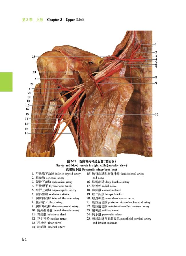
圖3-11 右腋窩內神經血管(前面觀) Nerves and blood vessels in right axilla(anterior view) 保留胸小肌 Pectoralis minor been kept54
圖3-12 右腋窩內臂叢和血管(前面觀) Nerves and blood vessels in right axilla(anterior view) 前壁**去除 Anterior wall been completely removed55
圖3-13 右腋淋巴結(前面觀) Lymph nodes in right axilla(anterior view)56
圖3-14 右臂前面肌、神經和血管(前面觀) Muscles, nerves and blood vessels in anterior compaial veins of right leg36
圖2-34 右小腿後區結構 Structures in posterior compartment of right leg37
圖2-35 右小腿前外側區和足背 Anterolateral compartment of right leg and dorsum of foot38
圖2-36 右小腿伸肌支持帶和腓骨肌支持帶 Right extensor retinaculum and peroneal retinaculum38
圖2-37 右足底結構(Ⅰ) Structures of sole of right foot(Ⅰ)39
圖2-38 右足底結構(Ⅱ) Structures of sole of right foot(Ⅱ)40
圖2-39 右踝管(下面觀) Right tarsal tunnel(inferior view)41
圖2-40 右足背結構 Structures of dorsum of right foot42
圖2-41 右小腿中部橫斷面(下面觀) Transverse section through middle of right leg(inferior view)43
第3章 上肢 Chapter 3 Upper Limb44(—72)
圖3-1 右鎖骨 Right clavicle45
圖3-2 右肩胛骨外側角 Lateral angle of right scapula45
圖3-3 右肩胛骨 Right scapula46
圖3-4 右肱骨 Right humerus47
圖3-5 右橈骨和尺骨 Right radius and ulna48
圖3-6 右手骨 Bones of right hand49
圖3-7 右肩關節 Right shoulder joint50
圖3-8 右肘關節 Right elbow joint51
圖3-9 左手骨關節 Joints of left hand52
圖3-10 右腋窩前壁 Anterior wall of right axilla53
圖3-11 右腋窩內神經血管(前面觀) Nerves and blood vessels in right axilla(anterior view) 保留胸小肌 Pectoralis minor been kept54
圖3-12 右腋窩內臂叢和血管(前面觀) Nerves and blood vessels in right axilla(anterior view) 前壁**去除 Anterior wall been completely removed55
圖3-13 右腋淋巴結(前面觀) Lymph nodes in right axilla(anterior view)56
圖3-14 右臂前面肌、神經和血管(前面觀) Muscles, nerves and blood vessels in anterior compartment of upper arm(anterior view)56
圖3-15 右肩胛區和臂後區的結構(後面觀) Structures of right scapular and posterior brachial regions(posterior view)57
圖3-16 右肩胛動脈網(後面觀) Right scapular anastomosis(posterior view)58
圖3-17 右肩袖肌(前面觀) Right rotator cuff(anterior view)59
圖3-18 右肩袖肌(後面觀) Right rotator cuff(posterior view)59
圖3-19 右肘窩和前臂皮神經和淺靜脈(前面觀) Cutaneous nerves and superficial veins on right cubital region and forearm(anterior view)60
圖3-20 右前臂前區的肌(前面觀) Muscles in anterior compartment of right forearm(anterior view)60
圖3-20 右前臂前區的肌(前面觀)(續)Muscles in anterior compartment of right forearm(anterior view)(continued)61
圖3-21 右肘窩內神經血管(前面觀) Nerves and blood vessels in right cubital fossa(anterior view)62
圖3-22 右前臂前區的神經血管(前面觀) Nerves and blood vessels in anterior compartment of right forearm(anterior view)63
圖3-23 右前臂旋前肌和旋後肌 Muscles for pronation and supination of right forearm64
圖3-24 右前臂後區肌(後面觀) Muscles in posterior compartment of right forearm(posterior view)65
圖3-25 右前臂後區神經血管(後面觀) Nerves and blood vessels in posterior compartment of right forearm(posterior view)65
圖3-26 右手掌腱膜(前面觀) Palmar aponeurosis of right hand(anterior view)66
圖3-27 右手掌淺弓和神經(前面觀) Superficial palmar arch and nerves of right hand(anterior view)66
圖3-28 右手掌深弓和神經(前面觀) Deep palmar arch and nerves of right hartment of upper arm(anterior view)56
圖3-15 右肩胛區和臂後區的結構(後面觀) Structures of right scapular and posterior brachial regions(posterior view)57
圖3-16 右肩胛動脈網(後面觀) Right scapular anastomosis(posterior view)58
圖3-17 右肩袖肌(前面觀) Right rotator cuff(anterior view)59
圖3-18 右肩袖肌(後面觀) Right rotator cuff(posterior view)59
圖3-19 右肘窩和前臂皮神經和淺靜脈(前面觀) Cutaneous nerves and superficial veins on right cubital region and forearm(anterior view)60
圖3-20 右前臂前區的肌(前面觀) Muscles in anterior compartment of right forearm(anterior view)60
圖3-20 右前臂前區的肌(前面觀)(續)Muscles in anterior compartment of right forearm(anterior view)(continued)61
圖3-21 右肘窩內神經血管(前面觀) Nerves and blood vessels in right cubital fossa(anterior view)62
圖3-22 右前臂前區的神經血管(前面觀) Nerves and blood vessels in anterior compartment of right forearm(anterior view)63
圖3-23 右前臂旋前肌和旋後肌 Muscles for pronation and supination of right forearm64
圖3-24 右前臂後區肌(後面觀) Muscles in posterior compartment of right forearm(posterior view)65
圖3-25 右前臂後區神經血管(後面觀) Nerves and blood vessels in posterior compartment of right forearm(posterior view)65
圖3-26 右手掌腱膜(前面觀) Palmar aponeurosis of right hand(anterior view)66
圖3-27 右手掌淺弓和神經(前面觀) Superficial palmar arch and nerves of right hand(anterior view)66
圖3-28 右手掌深弓和神經(前面觀) Deep palmar arch and nerves of right hand(anterior view)67
圖3-29 右手屈指肌腱和滑膜鞘(前面觀) Flexor tendons and synovial sheaths of right hand(anterior view)67
圖3-30 右手腕部神經血管(前面觀) Nerves and blood vessels of right wrist and hand(anterior view)68
圖3-31 右手腕部皮神經和淺靜脈(後面觀) Cutaneous nerves and superficial veins of right wrist and hand(posterior view)69
圖3-32 右手背伸肌腱和動脈(後面觀) Extensor tendons and arteries on dorsal aspect of right wrist and hand(posterior view)69
圖3-33 右手肌 Muscles of right hand70
圖3-34 右示指屈肌腱和腱紐(外側觀) Flexor tendons and vincula tendinum of index finger of right hand(lateral view)71
圖3-35 右臂中部橫斷面(下面觀) Transverse section through middle of right upper arm(inferior view)71
圖3-36 右前臂中部橫斷面(下面觀) Transverse section through middle of right forearm(inferior view)72
圖3-37 右手掌橫斷面(下面觀) Transverse section through metacarpals of right hand(inferior view)72
第4章 胸 Chapter 4 Thorax73(—100)
圖4-1 左側肋骨(上面觀) Left ribs(superior view)74
圖4-2 胸骨 Sternum74
圖4-3 肋與胸骨連接(前面觀) Joints of ribs and sternum(anterior view)75
圖4-4 胸前壁皮神經和淺靜脈(前面觀) Cutaneous nerves and superficial veins on anterior thoracic wall(anterior view)75
圖4-5 女性乳房(前面觀) Female breast(anterior view)76
圖4-6 胸前壁(內面觀) Anterior wall of thorax(internal view)76
圖4-7 胸膜與胸膜腔(前面觀) Pleura and pleural cavity(anterior view)77
圖4-8 右肺根和肺韌帶(前面觀) Root of right lung and pulmnd(anterior view)67
圖3-29 右手屈指肌腱和滑膜鞘(前面觀) Flexor tendons and synovial sheaths of right hand(anterior view)67
圖3-30 右手腕部神經血管(前面觀) Nerves and blood vessels of right wrist and hand(anterior view)68
圖3-31 右手腕部皮神經和淺靜脈(後面觀) Cutaneous nerves and superficial veins of right wrist and hand(posterior view)69
圖3-32 右手背伸肌腱和動脈(後面觀) Extensor tendons and arteries on dorsal aspect of right wrist and hand(posterior view)69
圖3-33 右手肌 Muscles of right hand70
圖3-34 右示指屈肌腱和腱紐(外側觀) Flexor tendons and vincula tendinum of index finger of right hand(lateral view)71
圖3-35 右臂中部橫斷面(下面觀) Transverse section through middle of right upper arm(inferior view)71
圖3-36 右前臂中部橫斷面(下面觀) Transverse section through middle of right forearm(inferior view)72
圖3-37 右手掌橫斷面(下面觀) Transverse section through metacarpals of right hand(inferior view)72
第4章 胸 Chapter 4 Thorax73(—100)
圖4-1 左側肋骨(上面觀) Left ribs(superior view)74
圖4-2 胸骨 Sternum74
圖4-3 肋與胸骨連接(前面觀) Joints of ribs and sternum(anterior view)75
圖4-4 胸前壁皮神經和淺靜脈(前面觀) Cutaneous nerves and superficial veins on anterior thoracic wall(anterior view)75
圖4-5 女性乳房(前面觀) Female breast(anterior view)76
圖4-6 胸前壁(內面觀) Anterior wall of thorax(internal view)76
圖4-7 胸膜與胸膜腔(前面觀) Pleura and pleural cavity(anterior view)77
圖4-8 右肺根和肺韌帶(前面觀) Root of right lung and pulmonary ligament(anterior view)78
圖4-9 肋間神經和血管(上面觀) Intercostal nerves and blood vessels(superior view)78
圖4-10 心於原位(前面觀) Heart in situ(anterior view)79
圖4-11 縱隔(左側面觀) Mediastinum(left lateral view)80
圖4-12 縱隔(右側面觀) Mediastinum(right lateral view)81
圖4-13 上腔靜脈、主動脈和肺動脈(前面觀) Superior vena cava, aorta and pulmonary arteries(anterior view)82
圖4-14 上縱隔和後縱隔(前面觀) Superior and posterior mediastinum(anterior view)83
圖4-15 奇靜脈和胸導管(右側面觀) Azygos vein and thoracic duct(right lateral view)84
圖4-16 漿膜心包(左側面觀) Serous pericardium(left lateral view)85
圖4-17 冠狀動脈和心的靜脈(右前外側面觀) Coronary arteries and cardiac veins(right anterolateral view)85
圖4-18 冠狀動脈和心的靜脈(左前側面觀) Coronary arteries and cardiac veins(left anterolateral view)86
圖4-19 冠狀動脈和心的靜脈(後面觀) Coronary arteries and cardiac veins(posterior view)86
圖4-20 右心室(已切開) Right ventricle of heart(dissected)87
圖4-21 左心室(已切開) Left ventricle of heart(dissected)88
圖4-22 心的瓣膜(上面觀) Valves of heart(superior view)89
圖4-23 心肌構築(前面觀) Myocardiac architecture(anterior view)90
圖4-24 冠狀動脈紅色鑄型 Red colored resin corrosion cast of the coronary arteries90
圖4-25 心傳導繫統 Cardiac conducting system91
圖4-26 喉、氣管及支氣管(前面觀) Larynx, trachea and bronchi(anterior view)91
圖4-27 支氣管肺段鑄型(後面觀) Colored resin corrosion cast of bronchopulmonary segments(posterior view)onary ligament(anterior view)78
圖4-9 肋間神經和血管(上面觀) Intercostal nerves and blood vessels(superior view)78
圖4-10 心於原位(前面觀) Heart in situ(anterior view)79
圖4-11 縱隔(左側面觀) Mediastinum(left lateral view)80
圖4-12 縱隔(右側面觀) Mediastinum(right lateral view)81
圖4-13 上腔靜脈、主動脈和肺動脈(前面觀) Superior vena cava, aorta and pulmonary arteries(anterior view)82
圖4-14 上縱隔和後縱隔(前面觀) Superior and posterior mediastinum(anterior view)83
圖4-15 奇靜脈和胸導管(右側面觀) Azygos vein and thoracic duct(right lateral view)84
圖4-16 漿膜心包(左側面觀) Serous pericardium(left lateral view)85
圖4-17 冠狀動脈和心的靜脈(右前外側面觀) Coronary arteries and cardiac veins(right anterolateral view)85
圖4-18 冠狀動脈和心的靜脈(左前側面觀) Coronary arteries and cardiac veins(left anterolateral view)86
圖4-19 冠狀動脈和心的靜脈(後面觀) Coronary arteries and cardiac veins(posterior view)86
圖4-20 右心室(已切開) Right ventricle of heart(dissected)87
圖4-21 左心室(已切開) Left ventricle of heart(dissected)88
圖4-22 心的瓣膜(上面觀) Valves of heart(superior view)89
圖4-23 心肌構築(前面觀) Myocardiac architecture(anterior view)90
圖4-24 冠狀動脈紅色鑄型 Red colored resin corrosion cast of the coronary arteries90
圖4-25 心傳導繫統 Cardiac conducting system91
圖4-26 喉、氣管及支氣管(前面觀) Larynx, trachea and bronchi(anterior view)91
圖4-27 支氣管肺段鑄型(後面觀) Colored resin corrosion cast of bronchopulmonary segments(posterior view)92
圖4-28 右肺 Right lung93
圖4-29 左肺 Left lung94
圖4-30 染料灌注示意左肺支氣管肺段 Colored territories for bronchopulmonary segments of left lung95
圖4-31 染料灌注示意右肺支氣管肺段 Colored territories for bronchopulmonary segments of right lung96
圖4-32 經肺動脈分叉胸部橫斷面(下面觀) Transverse section through thorax at level of bifurcation of pulmonary trunk(inferior view)97
圖4-33 經心長軸胸部斜橫斷面(上面觀) Oblique transverse section through thorax at long axis of heart(superior view)97
圖4-34 軀干正中矢狀斷面(左側面觀) Median sagittal section of trunk(left lateral view)98
圖4-35 經齒突頭、頸和軀干冠狀斷面(前面觀) Coronary section through head, neck and trunk at level of dens of axis(anterior view)99
圖4-36 膈(下面觀) Diaphragm(inferior view)100
圖4-37 膈(上面觀) Diaphragm(superior view)100
第5章 腹 Chapter 5 Abdomen101(—128)
圖5-1 腹前外側壁的淺靜脈(前面觀) Superficial veins on anterolateral wall of abdomen(anterior view)102
圖5-2 腹前外側壁的肌和腱膜(前面觀) Muscles and aponeurosis of anterolateral abdominal wall(anterior view)103
圖5-3 左側腹股溝管(前面觀) Left inguinal canal(anterior view)104
圖5-4 左側腹前外壁與精索的關繫(Ⅰ)(前面觀) Relationships of left half of anterolateral abdominal wall to spermatic cord(Ⅰ)(anterior view)105
圖5-4 左側腹前外壁與精索的關繫(Ⅱ)(前面觀) Relationships of left half of anterolateral abdominal wall to spermatic cord(Ⅱ)(anterior view)106
圖5-5 左側腹前外壁(後面觀) Left half of anterolateral a92
圖4-28 右肺 Right lung93
圖4-29 左肺 Left lung94
圖4-30 染料灌注示意左肺支氣管肺段 Colored territories for bronchopulmonary segments of left lung95
圖4-31 染料灌注示意右肺支氣管肺段 Colored territories for bronchopulmonary segments of right lung96
圖4-32 經肺動脈分叉胸部橫斷面(下面觀) Transverse section through thorax at level of bifurcation of pulmonary trunk(inferior view)97
圖4-33 經心長軸胸部斜橫斷面(上面觀) Oblique transverse section through thorax at long axis of heart(superior view)97
圖4-34 軀干正中矢狀斷面(左側面觀) Median sagittal section of trunk(left lateral view)98
圖4-35 經齒突頭、頸和軀干冠狀斷面(前面觀) Coronary section through head, neck and trunk at level of dens of axis(anterior view)99
圖4-36 膈(下面觀) Diaphragm(inferior view)100
圖4-37 膈(上面觀) Diaphragm(superior view)100
第5章 腹 Chapter 5 Abdomen101(—128)
圖5-1 腹前外側壁的淺靜脈(前面觀) Superficial veins on anterolateral wall of abdomen(anterior view)102
圖5-2 腹前外側壁的肌和腱膜(前面觀) Muscles and aponeurosis of anterolateral abdominal wall(anterior view)103
圖5-3 左側腹股溝管(前面觀) Left inguinal canal(anterior view)104
圖5-4 左側腹前外壁與精索的關繫(Ⅰ)(前面觀) Relationships of left half of anterolateral abdominal wall to spermatic cord(Ⅰ)(anterior view)105
圖5-4 左側腹前外壁與精索的關繫(Ⅱ)(前面觀) Relationships of left half of anterolateral abdominal wall to spermatic cord(Ⅱ)(anterior view)106
圖5-5 左側腹前外壁(後面觀) Left half of anterolateral abdominal wall(posterior view)107
圖5-6 腹腔內器官(前面觀) The organs in abdominal cavity(anterior view)108
圖5-7 網膜囊前壁(前面觀) Anterior wall of omenta bursa(anterior view)109
圖5-8 網膜囊後壁(沿胃大彎切開大網膜並向上翻起胃, 前面觀) Posterior wall of omenta bursa(greater omentum has been dissected and stomach has been raised upward, anterior view)110
圖5-9 胃的外形與分部(前面觀) External appearance and subdivision of stomach(anterior view)111
圖5-10 胃黏膜(胃前壁已切除,前面觀) Internal appearance of stomach(anterior wall of stomach has been removed, anterior view)111
圖5-11 胃小彎的神經血管(前面觀) Nerves and blood vessels along lesser curvature of stomach(anterior view)112
圖5-12 腹腔干及三個分支(前面觀) Celiac trunk and its three branches(anterior view)112
圖5-13 腹腔干與胰(前面觀) Celiac trunk and pancreas(anterior view)113
圖5-14 腸繫膜上動脈與胰頭(前面觀) Superior mesenteric artery and head of pancreas(anterior view)113
圖5-15 胰腺動脈和胰管的彩色鑄型(前面觀) Colored resin corrosion cast for arteries of pancreas and pancreatic duct(anterior view)114
圖5-16 脾 Spleen114
圖5-17 肝 Liver115
圖5-18 肝內血管和膽管(部分肝組織已被移除) Blood vessels and biliary ducts within liver(portions of substance of liver have been removed)116
圖5-19 肝外膽道和胰管(膽囊和十二指腸已被切開) Extrahepatic biliary ducts and pancreatic duct(gallbladder and duodenum have been opened)116
圖5-20 腸繫膜上動脈(前面觀) Superior mesenteric artery(antbdominal wall(posterior view)107
圖5-6 腹腔內器官(前面觀) The organs in abdominal cavity(anterior view)108
圖5-7 網膜囊前壁(前面觀) Anterior wall of omenta bursa(anterior view)109
圖5-8 網膜囊後壁(沿胃大彎切開大網膜並向上翻起胃, 前面觀) Posterior wall of omenta bursa(greater omentum has been dissected and stomach has been raised upward, anterior view)110
圖5-9 胃的外形與分部(前面觀) External appearance and subdivision of stomach(anterior view)111
圖5-10 胃黏膜(胃前壁已切除,前面觀) Internal appearance of stomach(anterior wall of stomach has been removed, anterior view)111
圖5-11 胃小彎的神經血管(前面觀) Nerves and blood vessels along lesser curvature of stomach(anterior view)112
圖5-12 腹腔干及三個分支(前面觀) Celiac trunk and its three branches(anterior view)112
圖5-13 腹腔干與胰(前面觀) Celiac trunk and pancreas(anterior view)113
圖5-14 腸繫膜上動脈與胰頭(前面觀) Superior mesenteric artery and head of pancreas(anterior view)113
圖5-15 胰腺動脈和胰管的彩色鑄型(前面觀) Colored resin corrosion cast for arteries of pancreas and pancreatic duct(anterior view)114
圖5-16 脾 Spleen114
圖5-17 肝 Liver115
圖5-18 肝內血管和膽管(部分肝組織已被移除) Blood vessels and biliary ducts within liver(portions of substance of liver have been removed)116
圖5-19 肝外膽道和胰管(膽囊和十二指腸已被切開) Extrahepatic biliary ducts and pancreatic duct(gallbladder and duodenum have been opened)116
圖5-20 腸繫膜上動脈(前面觀) Superior mesenteric artery(anterior view)117
圖5-21 回結腸動脈(前面觀) Ileocolic artery(anterior view)117
圖5-22 腸繫膜下動脈(前面觀) Inferior mesenteric artery(anterior view)118
圖5-23 回盲部(前面觀) Ileocecal junction(anterior view)118
圖5-24 肝門靜脈(空腸、回腸、橫結腸連同它們的繫膜已除去) Hepatic portal vein(jejunum, ileum, transverse colon and their mesentery have been removed)119
圖5-25 腹膜後隙的器官和血管(前面觀) Viscera and blood vessels within retroperitoneum(anterior view)120
圖5-26 腎冠狀切面 Coronary section of kidney121
圖5-27 腎內結構 Internal structures of kidney121
圖5-28 腎盞及腎盂 Renal calices and pelvis121
圖5-29 腎和腎上腺的動脈(前面觀) Arteries of kidneys and suprarenal glands(anterior view)122
圖5-30 副腎動脈及雙輸尿管(前面觀) Accessory renal artery and double ureter(anterior view)122
圖5-31 馬蹄腎(前面觀) Horseshoe kidney(anterior view)122
圖5-32 左腎被膜(前面觀) Coverings of left kidney(anterior view)123
圖5-33 腎後面毗鄰(後面觀) Structures related to posterior surface of kidney(posterior view)124
圖5-34 腰動脈和腰靜脈(右前外側觀) Lumbar arteries and veins(right anterolateral view)124
圖5-35 腹後隙的神經和血管(前面觀) Nerves and blood vessels within retroperitoneum(anterior view)125
圖5-36 腹後隙的神經(前面觀) Nerves within retroperitoneum(anterior view)126
圖5-37 腹後壁壁腹膜(前面觀) Posterior parietal peritoneum(anterior view)127
圖5-38 經幽門腹部橫斷面(下面觀) Transverse section through abdomen at level of pylorus(inferior view)128
第6erior view)117
圖5-21 回結腸動脈(前面觀) Ileocolic artery(anterior view)117
圖5-22 腸繫膜下動脈(前面觀) Inferior mesenteric artery(anterior view)118
圖5-23 回盲部(前面觀) Ileocecal junction(anterior view)118
圖5-24 肝門靜脈(空腸、回腸、橫結腸連同它們的繫膜已除去) Hepatic portal vein(jejunum, ileum, transverse colon and their mesentery have been removed)119
圖5-25 腹膜後隙的器官和血管(前面觀) Viscera and blood vessels within retroperitoneum(anterior view)120
圖5-26 腎冠狀切面 Coronary section of kidney121
圖5-27 腎內結構 Internal structures of kidney121
圖5-28 腎盞及腎盂 Renal calices and pelvis121
圖5-29 腎和腎上腺的動脈(前面觀) Arteries of kidneys and suprarenal glands(anterior view)122
圖5-30 副腎動脈及雙輸尿管(前面觀) Accessory renal artery and double ureter(anterior view)122
圖5-31 馬蹄腎(前面觀) Horseshoe kidney(anterior view)122
圖5-32 左腎被膜(前面觀) Coverings of left kidney(anterior view)123
圖5-33 腎後面毗鄰(後面觀) Structures related to posterior surface of kidney(posterior view)124
圖5-34 腰動脈和腰靜脈(右前外側觀) Lumbar arteries and veins(right anterolateral view)124
圖5-35 腹後隙的神經和血管(前面觀) Nerves and blood vessels within retroperitoneum(anterior view)125
圖5-36 腹後隙的神經(前面觀) Nerves within retroperitoneum(anterior view)126
圖5-37 腹後壁壁腹膜(前面觀) Posterior parietal peritoneum(anterior view)127
圖5-38 經幽門腹部橫斷面(下面觀) Transverse section through abdomen at level of pylorus(inferior view)128
第6
| | |
| | | | |
|