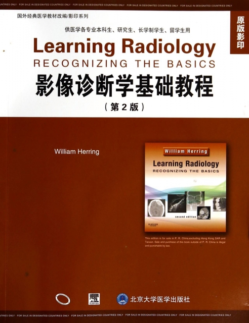
| | | | 影像診斷學基礎教程(供醫學各專業本科生研究生長學制學生留學生用原版影印第2版)/國外經典醫學教材改編影印繫列 | | 該商品所屬分類:醫學 -> 醫技學 | | 【市場價】 | 864-1252元 | | 【優惠價】 | 540-783元 | | 【介質】 | book | | 【ISBN】 | 9787565908026 | | 【折扣說明】 | 一次購物滿999元台幣免運費+贈品
一次購物滿2000元台幣95折+免運費+贈品
一次購物滿3000元台幣92折+免運費+贈品
一次購物滿4000元台幣88折+免運費+贈品
| | 【本期贈品】 | ①優質無紡布環保袋,做工棒!②品牌簽字筆 ③品牌手帕紙巾
|
|
| 版本 | 正版全新電子版PDF檔 | | 您已选择: | 正版全新 | 溫馨提示:如果有多種選項,請先選擇再點擊加入購物車。*. 電子圖書價格是0.69折,例如了得網價格是100元,電子書pdf的價格則是69元。
*. 購買電子書不支持貨到付款,購買時選擇atm或者超商、PayPal付款。付款後1-24小時內通過郵件傳輸給您。
*. 如果收到的電子書不滿意,可以聯絡我們退款。謝謝。 | | | |
| | 內容介紹 | |

-
出版社:北京大學醫學
-
ISBN:9787565908026
-
作者:(美)郝林
-
頁數:318
-
出版日期:2014-07-01
-
印刷日期:2014-07-01
-
包裝:平裝
-
開本:16開
-
版次:1
-
印次:1
-
字數:650千字
-
郝林編寫的《影像診斷學基礎教程(供醫學各專
業本科生研究生長學制學生留學生用原版影印第2版)
》為英文影像診斷學教材,由國際知名專家編寫,受
到世界各地讀者歡迎,並被世界多所著名醫學院校選
定為影像診斷學教材。從基礎、正常異常、胸部影像
開始繫統講解。知識精煉,解構精巧。有重點、要點
,以及診斷PITFULL等各種總結,方便學生抓住重點
。
-
Chapter 1
Recognizing Anything: An Introduction to Imaging
Modalities
Let There Be Light... and Dark, and Shades of Gray
Conventional Radiography (Plain Films)
Computed Tomography (CI or CAT Scans)
Ultrasound (US)
Magnetic Resonance imaging (MR )
Terminology
The Best System Is the Qne That Works
Conventions Used in This Book
Chapter 2
Recognizing Normal Chest Anatomy and a Technically
Adequate Chest Radiograph
The Normal Frontal Chest Radiograph
The Lateral Chest Radiograph
Five Key Areas on the Lateral Chest X-Ray
Evaluating the Chest Radiograph for Technical Adequacy
Chapter 3
Recognizing Airspace Versus Interstitial Lung
Disease
Classifying Parenchymal Lung Disease
Characteristics of Airspace Disease ]
Some Causes of Airspace Disease
Characteristics of Interstitial Lung Disease
Some Causes of Interstitial Lung Disease
Chapter 4
Recognizing the Causes of an Opacified
Hemithorax
Atelectasis of the Entire Lung
Massive Pleural Effusion
Pneumonia of an Entire Lung
Postpneumonectomy
Chapter 5
Recognizing Atelectasis
What Is Atelectasis?
Signs of Atelectasis
Types of Atelectasis
Patterns of Collapse in Lobar Atelectasis
How Atelectasis Resolves
Chapter 6
Recognizing a Pleural Effusion
Normal Anatomy and Physiology of the Pleural Space
Causes of Heural Effusions
Types of Pleural Effusions
Side Specificity of Pleural Effusions
Recognizing the Different Appearances of Pleural Effusions
Loculated Effusions
Chapter 7
Recognizing Pneumonia
General Considerations
General Characteristics of Pneumonia SO
Patterns of Pneumonia
Aspiration
Localizing Pneumonia SS
How Pneumonia Resolves
Chapter 8
Recognizing Pneumothorax, Pneumomediastinum,
Pneumopericardium, and Subcutaneous
Emphysema
Recognizing a Pneumothoraxg
Recognizing the Pitfalls in Overdiagnosing a Pneumothorax
Types of Pneumothoraces
Causes ofa Pneumothorax
Other Ways to Diagnose a Pneumothorax
Pulmonary Interstitial Emphysema
Recognizing Pneumomediastinum
Recognizing Pneumopericardium
Recognizing Subcutaneous Emphysema
Chapter 9
Recognizing Adult Heart Disease
Recognizing an Enlarged Cardiac Silhouette
Pericardial Effusion
Extracardiac Causes of Apparent Cardiac Enlargement
Effect of Projection on Perception of Heart Size
Identifying Cardiac Enlargement on an Anteroposterior Chest
Radiograph
Recognizing Cardiomegaly on the Lateral Chest Radiographg
Recognizing Cardiomegaly in Infants
Normal Cardiac Contours
Normal Pulmonary Vasculature
General Principles of Cardiac Imaging
Recognizing Common Cardiac Diseases
Chapter 10
Recognizing the Correct Placement of Lines and Tubes:
Critical Care Radiology
Endotracheal and TracheostomyTubes
Intravascular Catheters
Pulmonary Drainage Tubes (Chest Tubes, Thoracotomy Tubes)
Cardiac Devices
Gastrointestinal Tubes and Lines
Chapter 11
Computed Tomography: Understanding the Basics and
Recognizing Normal Anatomy
Introduction to CT
Intravenous Contrast in CT Scanning
Oral Contrast in CTScanningg
Normal Chest CT Anatomy
Cardiac CT
Abdominal CT
Chapter 12
Recognizing Diseases of the Chest
Mediastinal Masses
Anterior Mediastinum g
Middle Mediastinum ]
Posterior Mediastinum ]
Solitary Nodule/Mass in the Lung
Bronchogenic Carcinoma
Metastatic Neoplasms in the Lung t
PulmonaryThromboembolic Disease
Chronic Obstructive Pulmonary Disease
BIebs and Bullae, Cysts and Cavities
Bronchiectasis
Chapter 13
Recognizing the Normal Abdomen: Conventional
Radiographs
What to Look For
Normal Bowel Gas Pattern
Normal Fluid Levels
Differentiating Large from Small Bowel
Acute Abdominal Series: The Views and What They Show
Calcifications
Organomegaly
Chapter 14
Recognizing Bowel Obstruction and Ileus
Abnormal Gas Patterns
Laws of the Gut
Functional Iteus, Localized: Sentinel Loops
Functional Ileus, Generalized: Adynamic Ileus
Mechanical Obstruction: Smalt Bowel Qbstruction (SBQ)
Mechanical Obstruction: Large Bowel Obstruction (LBO)
Volvulus of the Colon
Intestinal Pseudo-obstruction (Ogilvie Syndrome)
Chapter 15
Recognizing Extraluminal Air in the Abdomen
Signs of Free intraperitoneal Air
Causes of Free Air
Signs of Extraperitoneal Air (Retroperitoneal Air)
Causes of Extraperitoneal Air
Signs of Air in the Bowel Wall
Causes and Significance of Air in the Bowel Wall
Signs of Air in the Biliary System
Causes of Air in the Biliary System
Chapter 16
Recognizing Abnormal Calcifications and
Their Causes
Patterns of Calcification
Rimlike Calcification
Linear or Tracklike Calcification
LameFar or Laminar Calcification
Cloudlike, Amorphous, or Popcorn Calcification
Location of Calcification
Chaptert 17
Recognizing the Imaging Findings of Trauma
Chest Trauma
Aortic Trauma
Abdominal Trauma
Pelvic Trauma
Chapter 18
Recognizing Gastrointestinal, Hepatic, and Urinary
Tract Abnormalities
Barium Studies of the Gastrointestinal Tract
Esophagus
Stomach and Duodenum
Small and Large Bowel
Pancreas
Hepatobiliary Abnormalities
Urinary Tract
Pelvis
Urinary Bladder
Chapter 19
Ultrasonography: Understanding the Principles and
Recognizing Normal and Abnormal Findings
How it Works
Doppler Ultrasonography
Adverse Effects and Safety Issues
Medical Uses of Ultrasonography
Biliary System
Urinary Tract
Abdominal Aortic Aneurysms
Female Pelvic Organs
Appendicitis
Pregnancy
Vascular Ultrasound
Deep Venous Thrombosis
Chapter 20
Magnetic Resonance Imaging: Understanding the
Principles and Recognizing the Basics
DANIEL J. KOWAL
How MRI Works
Hardware That Makes Up an MRI Scanner
What Happens Once Scanning Begins
Pulse Sequences
How Can You Identify a Tl-Weighted or T -Weighted Image?
MRI Contrast: General Considerations
MRI Safety Issues
Diagnostic Applications of MR/
Chapter 21
Recognizing Abnormalities of Bone Density
Normal Bone Anatomy
The Effect of Bone Physiology on Bone Anatomy
Recognizing a Generalized Increase in Bone Density
Recognizing a Focal Increase in Bone Density
Recognizing a Generalized Decrease in Bone Density
Recognizing a Focal Decrease in Bone Density
Pathologic Fractures
Chapter 22
Recognizing Fractures and Dislocations
Recognizing an Acute Fracture
Recognizing Dislocations and Subluxations
Describing Fractures
Avulsion Fractures
Salter-Harris Fractures: Epiphyseal Plate Fractures in Children
Child Abuse
Stress Fractures
Common Fracture Eponyms
Some Easily Missed Fractures or Dislocations
Fracture Healing
Chapter 23
Recognizing Joint Disease: An Approach to
Arthritis
Anatomy of a Joint
Classification of Arthritis
Hypertrophic Arthritis
Erosive Arthritis
Infectious Arthritis
Chapter 24
Recognizing Some Common Causes of Neck and
Back Pain
Conventional Radiology, MRI, and
The Normal Spine
Back Pain
Herniated Disks
Degenerative Disk Disease
Osteoarthritis of the Facet Joints
Diffuse Idiopathic Skeletal Hyperostosis
Compression Fractures of the Spine
Spondylolisthesis and Spondylolysis
Spinal Stenosis
Malignancy Involving the Spine
MRI in Metastatic Spine Disease
Infections of the Spine: Diskitis and Osteomyelitis
Spinal Trauma
Chapter 25
Recognizing Some Common Causes of Intracranial
Pathology
Normal Anatomy
MRI and the Brain
Head Trauma
Intracranial Hemorrhage
Diffuse Axonal Injury
Increased Intracranial Pressure
Stroke
Ruptured Aneurysms
Hydrocephalus
Cerebral Atrophy
Brain Tumors
Multiple Sclerosis
Terminology
Appendix: Recognizing What to Order
Bibliography
Last Printed Paqe
| | |
| | | | |
|


